Clinical Use of the Vetario T50M Incubator in Guinea Pig Dental Aftercare
Dental disease is one of the most common clinical presentations in guinea pigs (Cavia porcellus), largely due to their continuously growing teeth and susceptibility to dietary and conformational imbalances. Affected individuals frequently present with weight loss, dysphagia, ptyalism, and reduced faecal output, reflecting both oral discomfort and secondary gastrointestinal dysfunction. Effective peri-anaesthetic management is therefore essential, as guinea pigs are particularly prone to hypothermia, ileus, and delayed recovery following sedation or general anaesthesia.
A three-year-old guinea pig was presented with a history of progressive weight loss, reduced appetite, and difficulty prehending and chewing food. Clinical examination raised suspicion of dental disease, and oral assessment under light sedation confirmed elongation of the cheek teeth with associated mucosal ulceration (Figure 1). Corrective dental burring was performed to restore occlusion and alleviate soft tissue trauma.
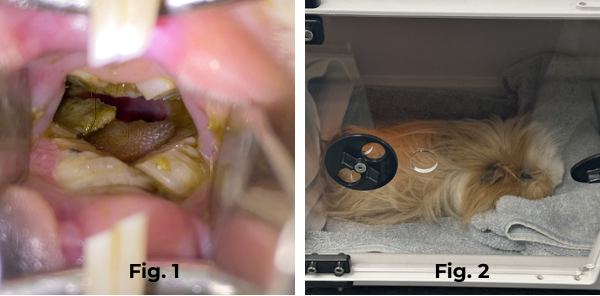
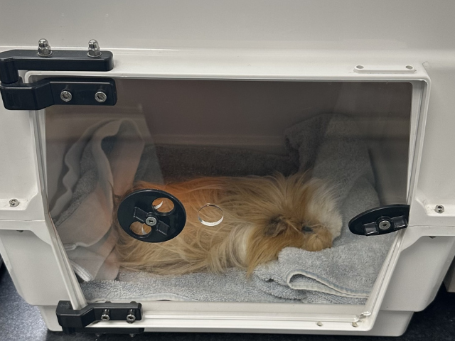
Given the recognised anaesthetic sensitivity of this species, particular emphasis was placed on post-operative care. Following the procedure, the patient was recovered in a Vetario incubator to provide a stable and controlled environment (Figure 2). The unit allowed precise thermal regulation, reducing the risk of post-anaesthetic hypothermia, which can significantly delay recovery and exacerbate gastrointestinal stasis. Supplemental oxygen was provided within the chamber to support cardiorespiratory function during the immediate recovery phase.
The enclosed, low-stimulus environment also played an important role in minimising stress, which is known to negatively impact recovery in prey species. Continuous visual monitoring through the transparent chamber enabled close observation of respiratory effort, posture, and behaviour without the need for repeated handling.
The guinea pig recovered smoothly, regaining normal posture and responsiveness within a short period. Early nutritional support was prioritised, with assisted syringe feeding initiated until voluntary food intake resumed. This intervention was critical in maintaining gastrointestinal motility and preventing ileus. Faecal output improved progressively over the following hours, indicating restoration of gut function.
The use of the incubator was central to the successful outcome in this case. Its ability to provide consistent warmth, facilitate oxygen supplementation, and create a calm recovery environment significantly enhanced patient stability. In small mammal practice, where peri-anaesthetic risks are considerable, such supportive care is essential in promoting safe recovery and optimising clinical outcomes.
Figure 1: Oral examination under light sedation demonstrating elongation of the cheek teeth with associated mucosal ulceration in a guinea pig (Cavia porcellus).
Figure 2: Post-operative recovery of the guinea pig within a controlled incubator environment, providing thermal support and oxygen supplementation following dental treatment.
Dr Sonya Miles.
BVSc CertAVP(ZM), CertAqV, MRCVS. RCVS Recognised Advanced Practitioner in Zoological Medicine. WAVMA Certified Aquatic Species Vet.
Just Exotics www.justexotics.co.uk